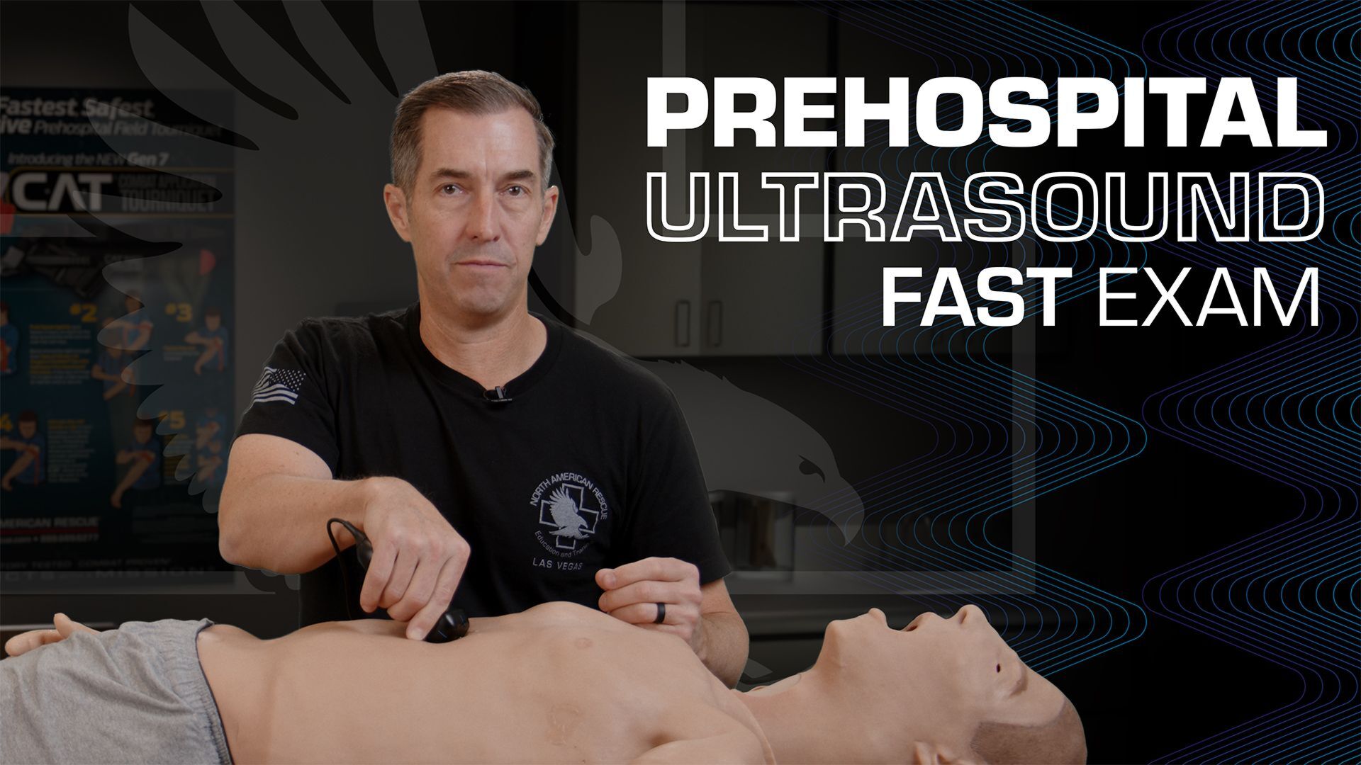

Hey everyone, doc Miles at North American Rescue. Today I'm going over some of the uses of ultrasound in the pre-hospital setting with your patients. So one of the most common uses and well-known uses is doing the FAST exam or the eFAST exam.
FAST Exam
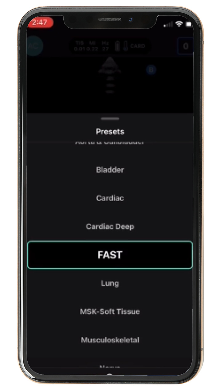
Very simple exam to learn. Takes a few practice runs, and then you're able to identify hemorrhage within the abdomen. So I'm going to focus on the abdominal portion of the FAST exam today. We're going to make sure that we've got our probe connected. Takes about two seconds to connect. We'll go to presets, and I'm going to put it on FAST. Make sure I've got gel on mine.
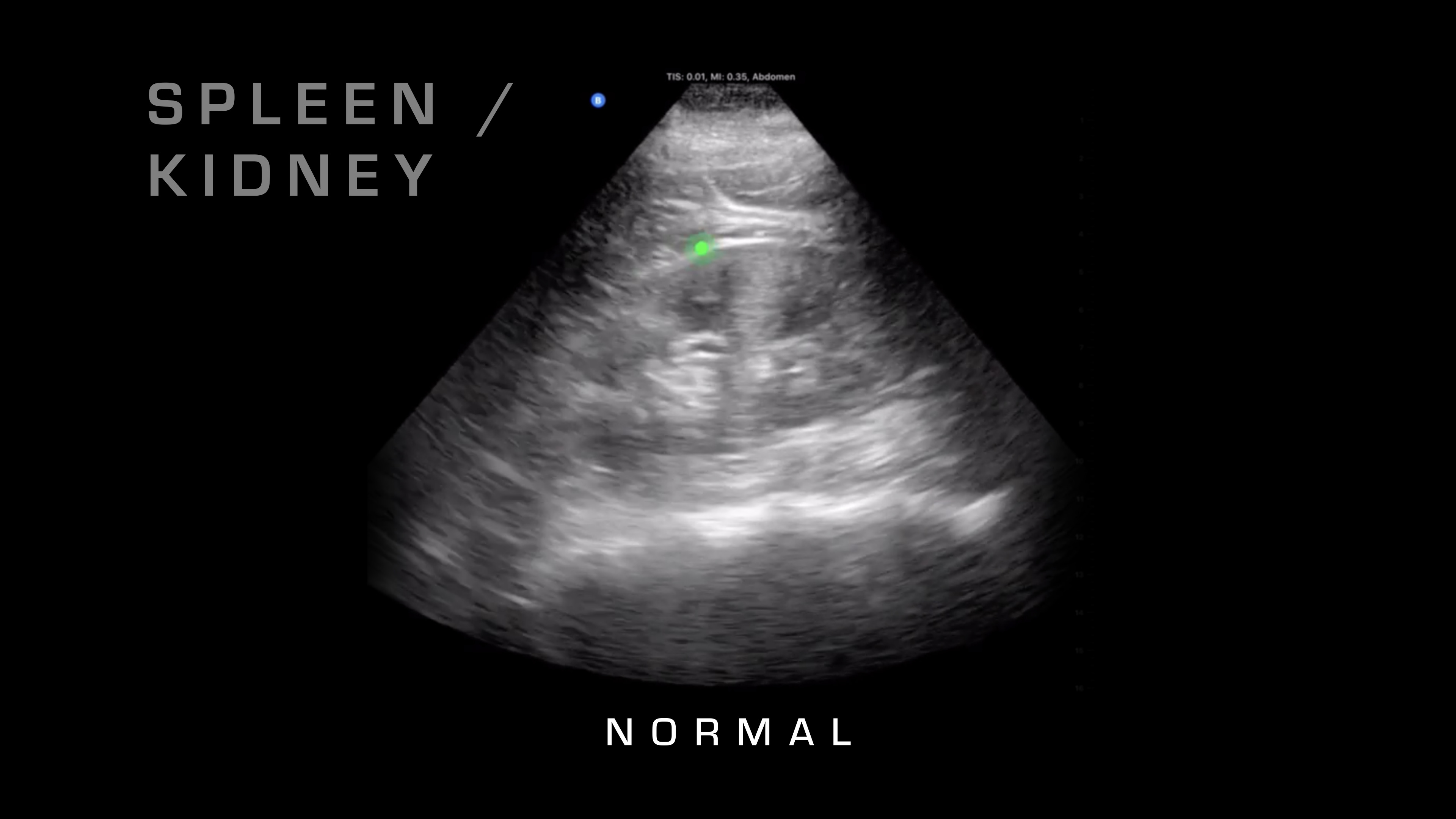
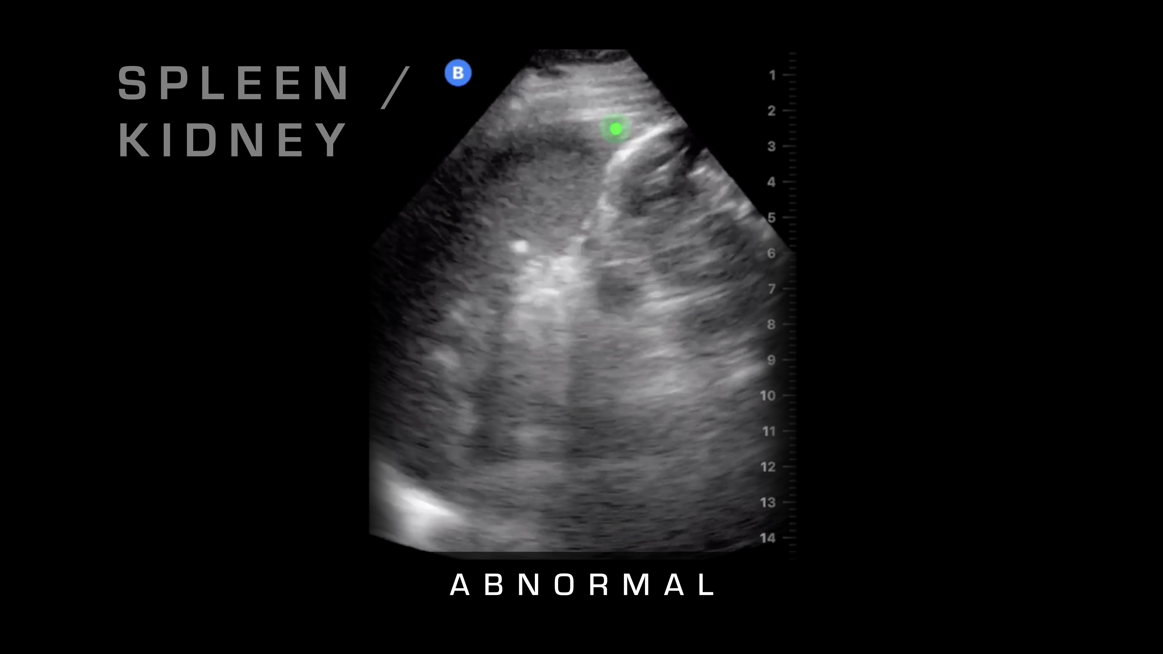
Spleen and Kidney
Then the first thing that I'm going to do is I'm going to place it down over here. What am I looking for here? I'm looking for the spleen and the kidney where they intersect. Sometimes you can tell if it's a cracked or a fractured spleen, if there's a big hematoma within the spleen, but really, we're looking for free fluid in the abdomen, which is going to be blood.
So what does blood in the abdomen look like? Blood looks like a black spot because, as the ultrasound waves penetrate through, it travels with minimal reflection, so it's just going to look like a black spot.
So I'll show you some normal, and then here's an abnormal one where you can see the fluid that's built up between the spleen and the kidney.
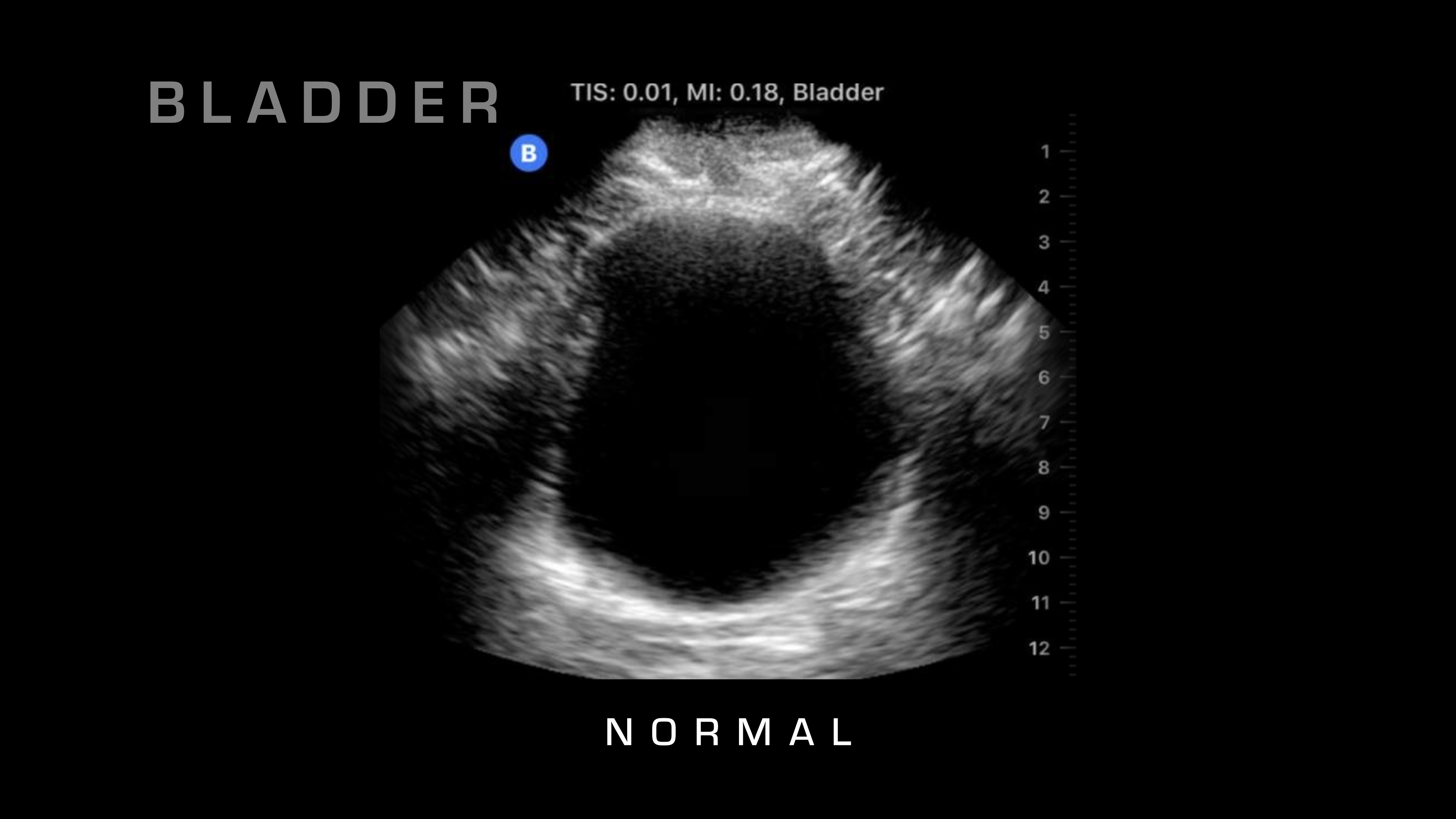
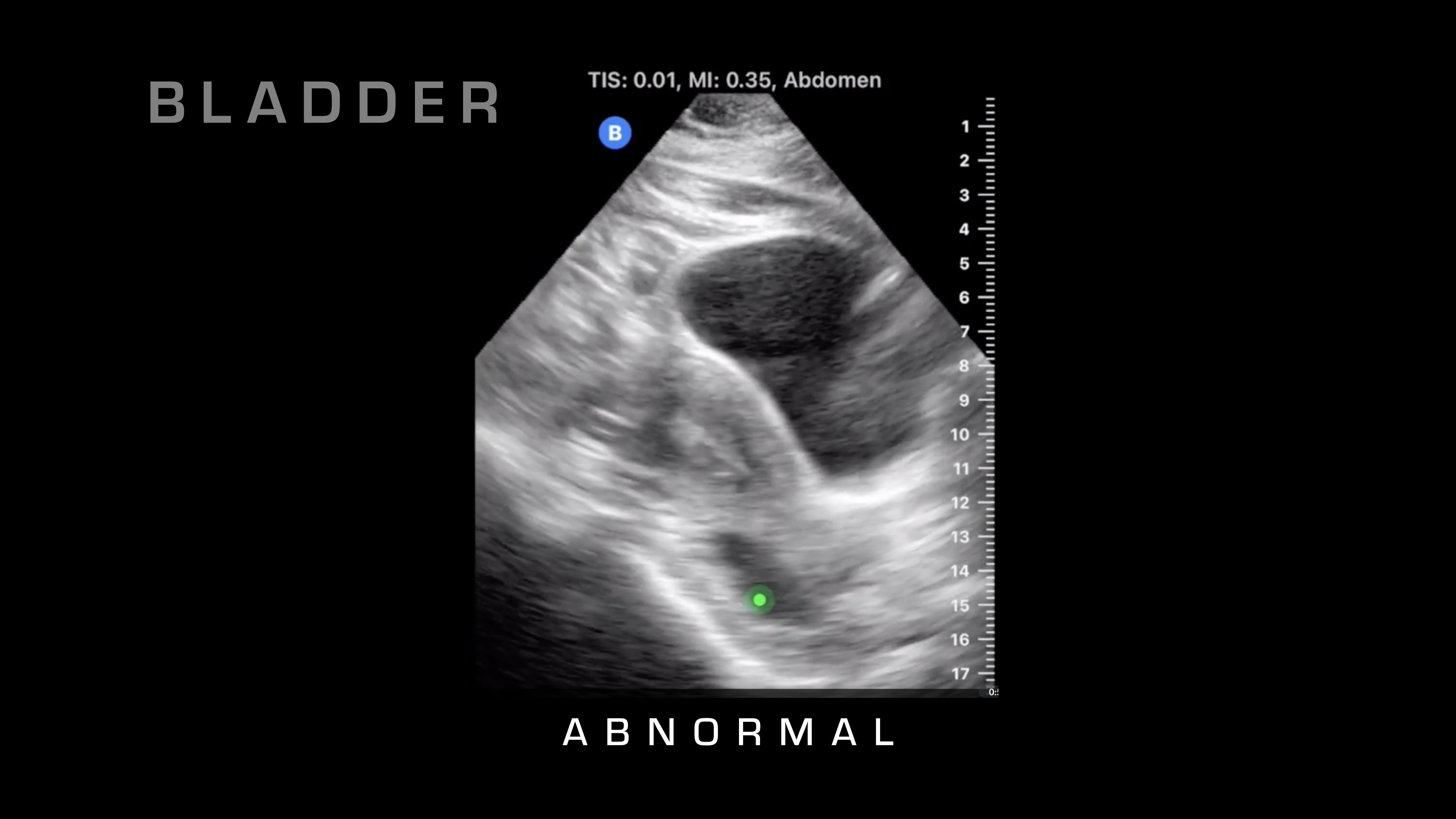
Pubic Symphysis
The next spot that I'm going to go to is it's going to be super pubic, so I'm going to go just over the pubic symphysis, and I'm going to angle basically straight down. What we're looking at here is we're going to see a large, fluid-filled structure that's going to be the bladder. We're looking for blood that is surrounding that, so either on the sides or if we penetrate all the way through the bladder, then we can take a look.
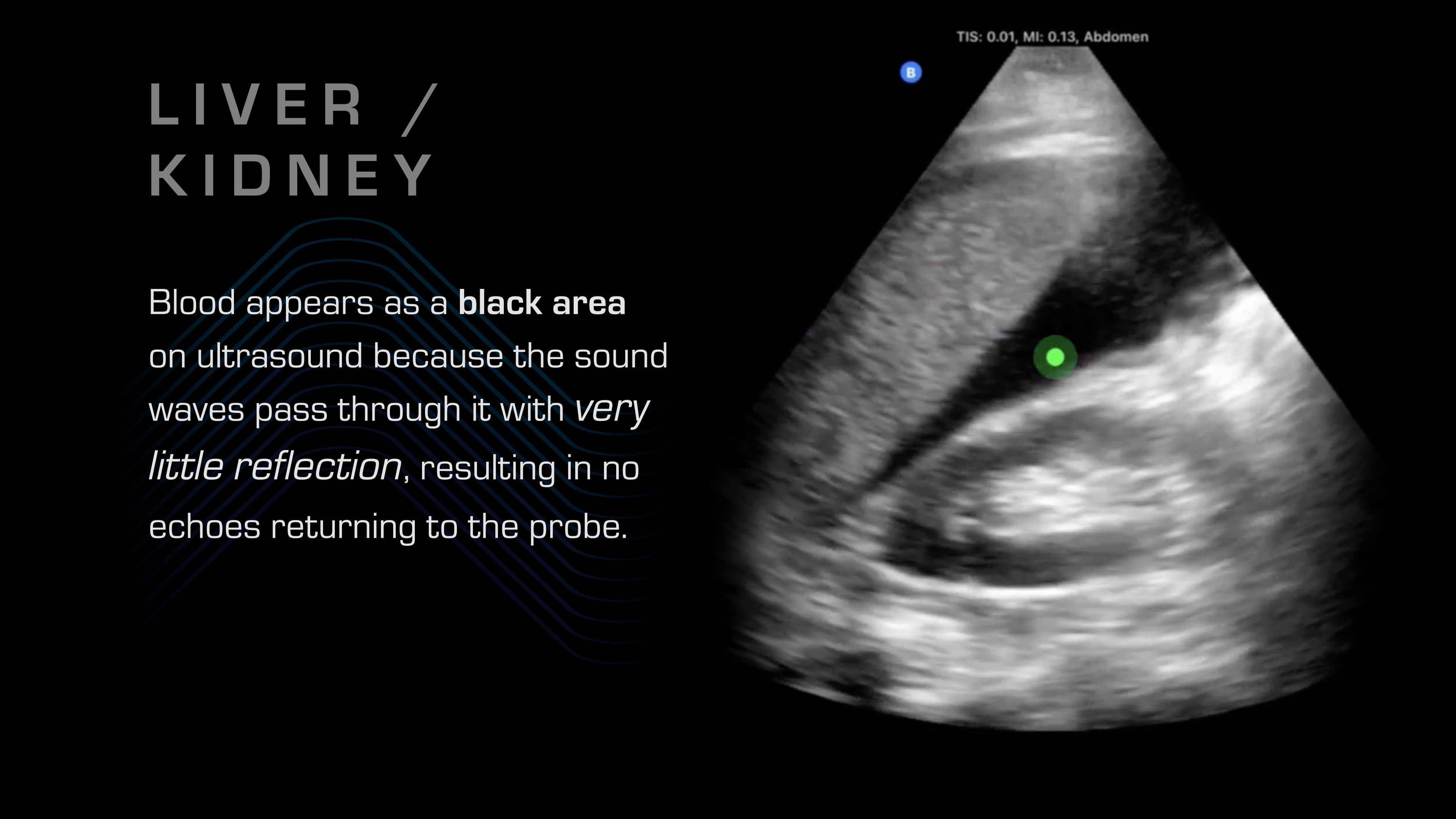
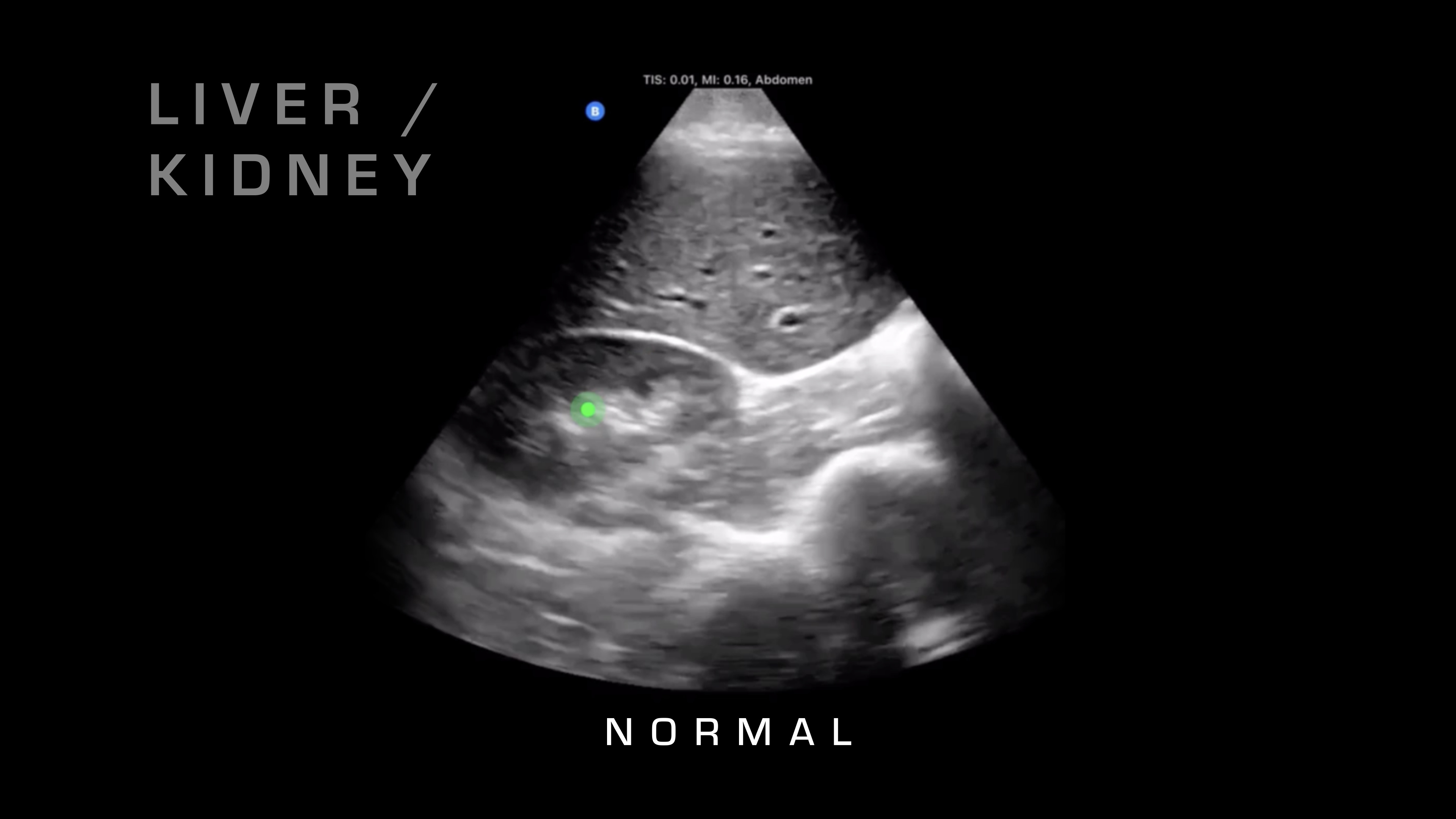
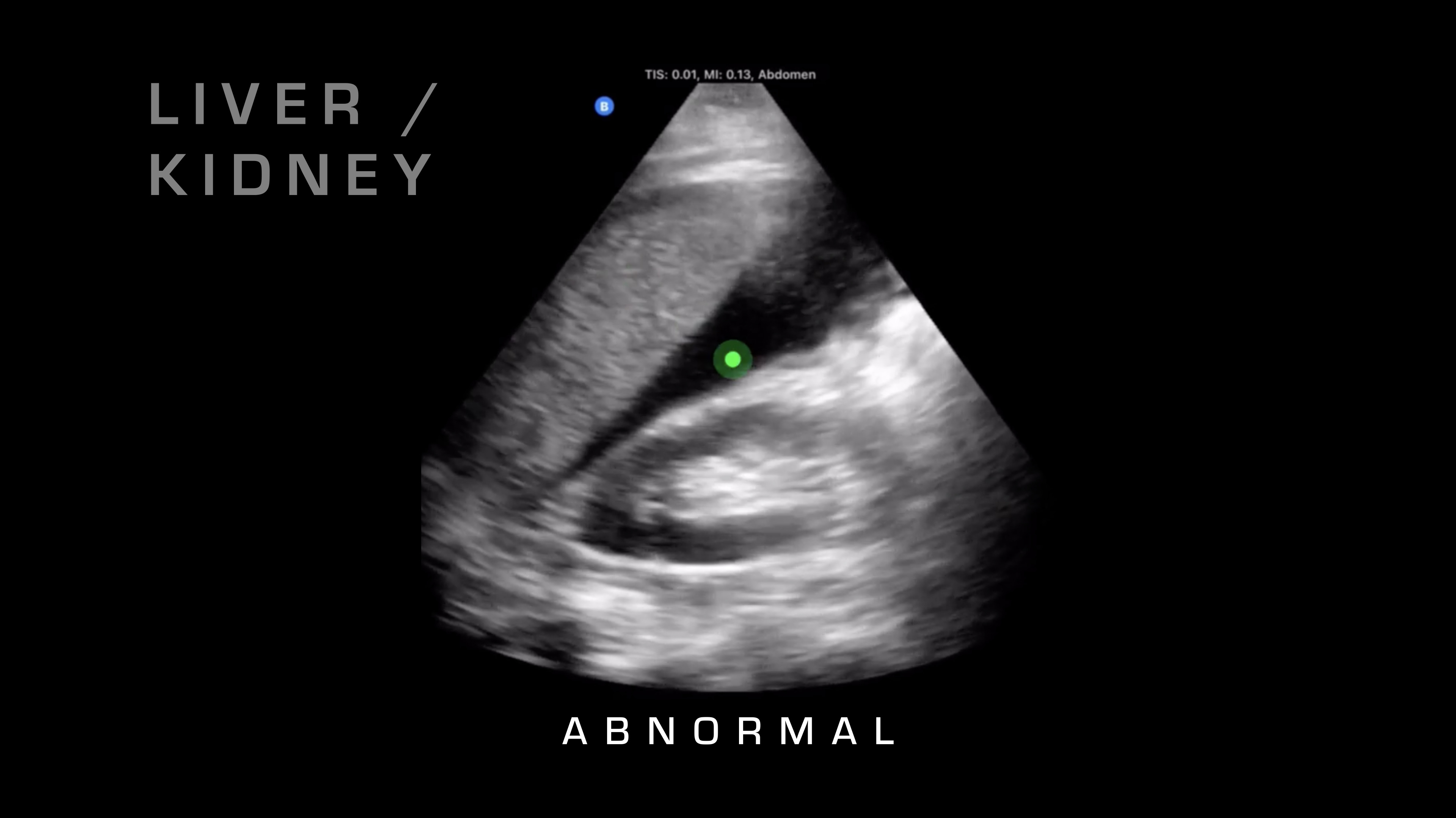
Liver & Kidney
If I want to go superficial, I'll swipe it down. If I want to go deeper, I'm going to swipe up and take a look. Next up, I'm going to go over on the right side of the patient, and what I'm going to be looking for is the liver and where that interfaces with the kidney. So there's a potential space there that shouldn't have any fluid between it, and if you see fluid between it, as you can see on this video here, then that's blood in the abdomen. So you know your patient is bleeding internally. That's obviously a risk for hemorrhagic shock, and that would warrant resuscitative care and rapid evacuation.
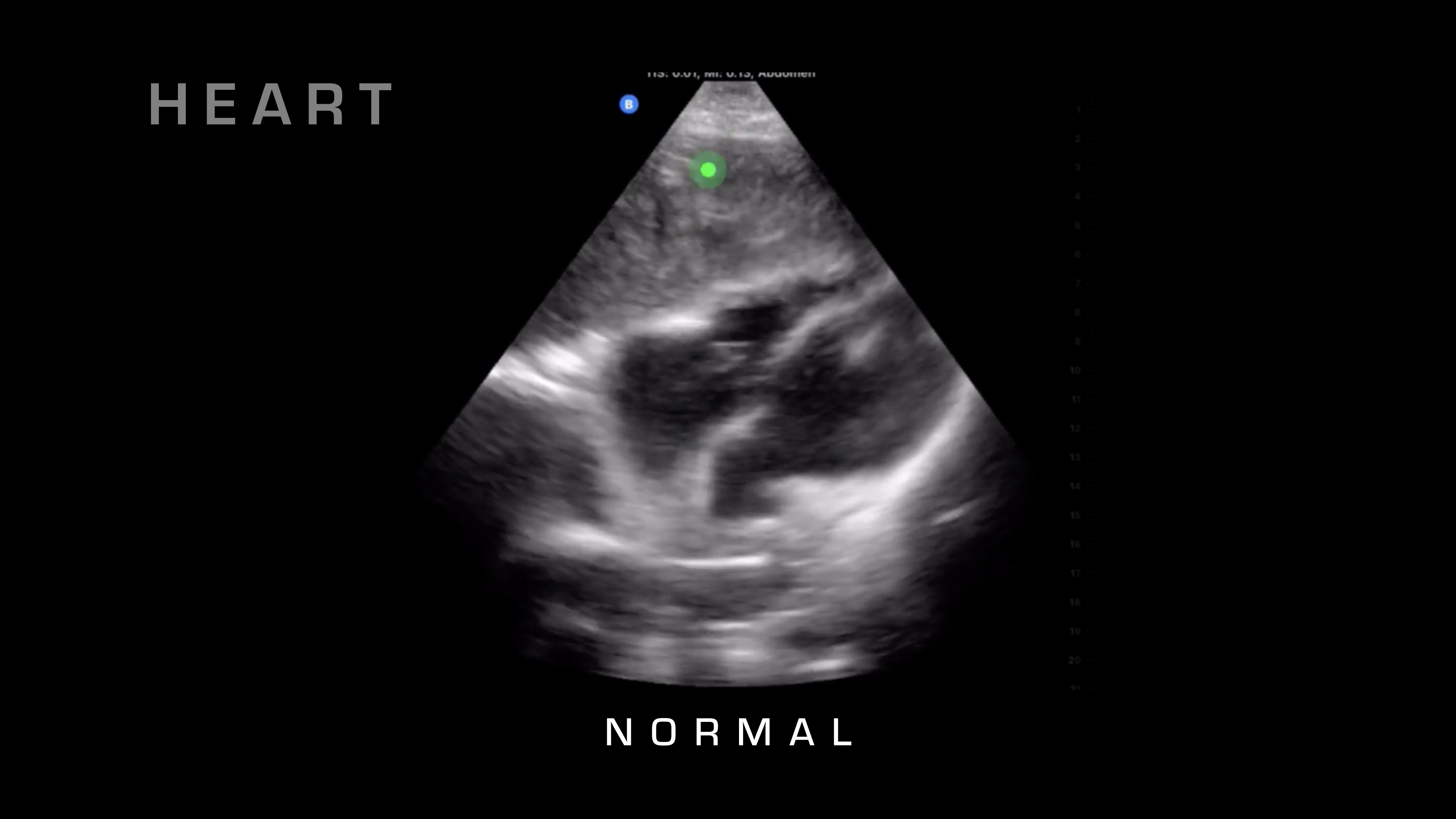
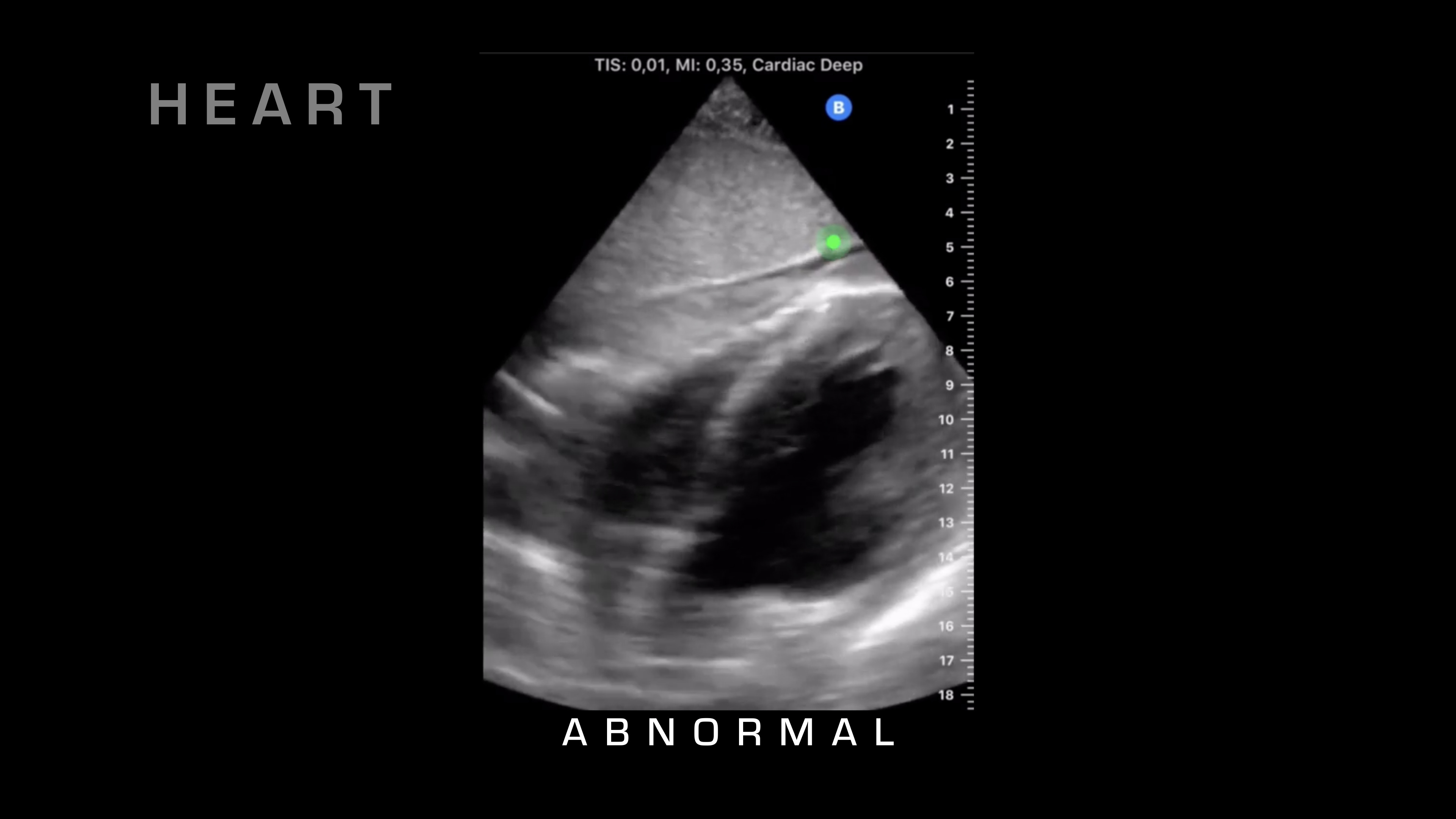
Cardiac Tamponade
So next up on our FAST exam, what we're going to do is we're going to take a look for cardiac tamponade, and we can also get a nice view of cardiac activity. So for cardiac tamponade, what I'm looking at is the heart beating. I'm looking for that sac surrounding the heart, and then if there's fluid in between those two, typically there shouldn't be any fluid between those two, but in cardiac tamponade, fluid will go in there, blood will go into that cavity, and it'll build up and build up, then restricting the heart and reducing cardiac output.
So there's a lot more tutorials. There are classes that you can take, you can do online education. The biggest thing is to just get one of these probes in your hand and start scanning people. The more you see normal, the more you're going to be able to recognize quickly what abnormal looks like. So hopefully that helps out on doing a FAST exam for you and showing you some of the things that you're going to be looking for.